Retina MCQs & Guide
600+ questions with detailed explanations. Master retina for FRCOphth, ICO, FAICO, DNB and NEET-SS.
Retina overview
Retina is the highest-weighted single subspecialty across modern ophthalmology exams — typically 18–25% of MCQs in FRCOphth Part 2, FICO and NEET-SS. The field has accelerated with anti-VEGF therapy, OCT, OCTA and gene therapy.
Clinical & exam importance
Diabetic retinopathy and AMD account for the bulk of blindness in working-age and elderly populations respectively. Retinal detachment is a surgical emergency. NEET-SS rankers are made or broken on the retina paper.
At a glance
- MCQ bank
- 600+
- Glossary terms
- 14+
- Free sample MCQs
- 25
- Matched products
- 2
Core anatomy & physiology refresher
The foundations you need before tackling clinical and surgical MCQs.
- Ten retinal layers; inner retina supplied by central retinal artery, outer retina by choriocapillaris via Bruch membrane.
- Macula is the central 5.5 mm, fovea 1.5 mm, foveola 0.35 mm — cone-only avascular zone.
- RPE has six key functions: phagocytosis, visual cycle, blood-retinal barrier, melanin absorption, growth factor secretion, ion transport.
- Bruch membrane: five layers between RPE and choriocapillaris; thickens with age, central to AMD.
- Vitreous is 99% water with hyaluronic acid and type II collagen; PVD typically by age 65.
High-yield retina facts for MCQs
The facts that appear most often in real exam stems. Learn them cold.
- 1Diabetic retinopathy staging (ETDRS): mild/moderate/severe NPDR, then PDR.
- 2Severe NPDR rule: 4 quadrants of haemorrhages, 2 quadrants venous beading, 1 quadrant IRMA (4-2-1 rule).
- 3PDR features: NVD, NVE, vitreous haemorrhage, tractional retinal detachment.
- 4DMO is the leading cause of vision loss in diabetic patients; treat with anti-VEGF +/- focal laser.
- 5AMD: dry (drusen, geographic atrophy) vs wet (CNV). Anti-VEGF for wet AMD.
- 6Rhegmatogenous RD: PVD with break, photopsia and floaters, curtain-like field loss.
- 7Macular hole stages (Gass): 1 (impending) → 2 (small full thickness) → 3 (>400 µm) → 4 (with PVD).
- 8CRVO: blood and thunder fundus, 4 quadrants of haemorrhage; ischaemic vs non-ischaemic by RAPD and FFA.
- 9BRVO most commonly at superotemporal AV crossings; treat macular oedema with anti-VEGF.
- 10Retinitis pigmentosa: triad of bone-spicule pigmentation, attenuated arterioles, waxy disc pallor.
- 11Best disease: vitelliform lesion with reduced Arden ratio on EOG; BEST1/VMD2 gene.
- 12Stargardt: pisciform flecks, dark choroid on FFA, ABCA4 gene.
- 13Central serous retinopathy: young men, type A personality, steroids; treat conservatively first.
- 14OCT layers to identify: ILM, NFL, GCL, IPL, INL, OPL, ONL, ELM, EZ, IZ, RPE/Bruch.
- 15OCTA detects flow without dye; useful in CNV, ischaemic maculopathy and capillary dropout in DR.
Related retina glossary terms
Exam-ready definitions filtered by subspecialty.
Matched products for Retina
Resources hand-picked for this subspecialty.
Or buy the complete bundle for your exam
These exam bundles include retina plus all the other topics you need.
FAICO MCQs
AIOS Fellowship exam MCQs mapped topic-by-topic to the FAICO syllabus.
Retina Deciphered
150+ pages of handwritten exam-ready notes covering the entire retina syllabus.

Glaucoma Exam Ready Notes
Complete glaucoma notes from pathophysiology to surgical management.

What's Included
- ✓1500+ FAICO-mapped MCQs
- ✓Topic-wise detailed explanations
- ✓Handwritten subspecialty notes
- ✓Mobile app access
- ✓Regular syllabus updates
Covered Topics
- ✓Retina
- ✓Glaucoma
- ✓Cornea
- ✓Cataract
- ✓Neuro-Ophthalmology
- ✓Uveitis
Instant access after purchase. Available on Android & iOS app.
Ophthal MCQs High Yield
High-yield MCQ bank across all ophthalmology subspecialties for rapid revision.
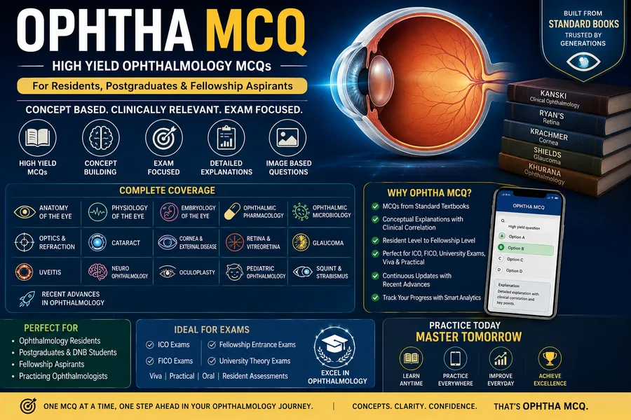
High Yield MCQs
1,000+ high-yield MCQs across all subspecialties with detailed explanations.
Eyelids Exam Ready Notes
Oculoplastics and eyelid disorders for your PG and fellowship exams.
What's Included
- ✓1000+ high-yield MCQs
- ✓Concise explanations
- ✓Quick revision notes
- ✓Mobile app access
- ✓Lifetime updates
Covered Topics
- ✓Cornea
- ✓Glaucoma
- ✓Retina
- ✓Neuro-Ophthalmology
- ✓Uveitis
- ✓Optics & Refraction
Instant access after purchase. Available on Android & iOS app.
General Ophthalmology
A mixed sampler across all ophthalmology subspecialties to test your overall readiness.
High Yield MCQs
1,000+ high-yield MCQs across all subspecialties with detailed explanations.
Retina Deciphered
150+ pages of handwritten exam-ready notes covering the entire retina syllabus.
Glaucoma Exam Ready Notes
Complete glaucoma notes from pathophysiology to surgical management.
ICO/FICO Past Papers
Last 10 years of ICO/FICO past papers with full explanations and answer keys.
What's Included
- ✓2000+ mixed ophthalmology MCQs
- ✓Cross-topic explanations
- ✓Combined notes and past papers
- ✓Mobile app access
- ✓Lifetime updates
Covered Topics
- ✓Anatomy
- ✓Optics & Refraction
- ✓Cornea
- ✓Lens & Cataract
- ✓Glaucoma
- ✓Retina
Instant access after purchase. Available on Android & iOS app.
Suggested study sequence
Six phases used by recent passers. Spread across 4–8 weeks per subspecialty.
- 1
1. Anatomy and imaging
OCT layers, FFA phases, OCTA flow slabs, autofluorescence.
- 2
2. Vascular retina
DR, RVO, hypertensive retinopathy, retinal artery occlusion.
- 3
3. Macular disease
AMD, macular hole, ERM, CSR, DMO.
- 4
4. Retinal detachment
Rheg, tractional, exudative. Surgical approaches: SB, PPV, PR.
- 5
5. Inherited retinal disease
RP, Stargardt, Best, X-linked retinoschisis, choroideremia.
- 6
6. Tumours and trauma
Retinoblastoma, choroidal melanoma, commotio retinae, choroidal rupture.
25 free retina MCQs with explanations
Exam-pattern stems with the correct option marked and the reasoning spelled out. Open a question to check your answer.
Q1.Earliest sign of diabetic retinopathy is:
Explanation: Microaneurysms (small saccular outpouchings of capillaries) are the earliest visible sign of non-proliferative diabetic retinopathy.
Q2.High-risk PDR per ETDRS criteria includes NVD ≥1/4–1/3 disc area OR:
Explanation: High-risk PDR: NVD ≥1/4–1/3 disc area, OR any NVD with VH/preretinal haemorrhage, OR NVE ≥1/2 disc area with VH/preretinal haemorrhage.
Q3.First-line treatment for centre-involving DME with vision 6/12 or worse:
Explanation: Per DRCR.net Protocols T/V, intravitreal anti-VEGF is first-line for centre-involving DME with decreased vision. Laser remains an option for selected non-centre or refractory eyes.
Q4.Sudden painless monocular loss of vision with a cherry-red spot is most consistent with:
Explanation: CRAO causes acute painless severe vision loss with a pale ischemic retina and cherry-red spot (intact foveal choroidal perfusion).
Q5.Cherry-red spot in a child with hepatosplenomegaly suggests:
Explanation: Cherry-red spot in storage disorders: Tay-Sachs (no organomegaly classically), Sandhoff, Niemann-Pick. With hepatosplenomegaly think Niemann-Pick / Sandhoff; among options Tay-Sachs is the classic association.
Q6.Roth spots are:
Explanation: Roth spots are flame-shaped haemorrhages with central pale (white) areas. Causes: endocarditis, leukemia, severe anaemia, diabetes, HIV.
Q7.Most useful imaging for an early macular hole:
Explanation: SD-OCT shows the foveal architecture and is the gold standard to stage and follow macular holes (Gass / IVTS classifications).
Q8.A 60-year-old with sudden flashes, floaters, and a curtain of vision loss most likely has:
Explanation: Flashes (photopsia) and a sudden shower of floaters in an older adult should prompt urgent retinal examination to rule out PVD with retinal tear or rhegmatogenous detachment.
Q9.Most common type of age-related macular degeneration:
Explanation: Dry AMD (drusen ± geographic atrophy) accounts for ~85-90% of cases; wet AMD causes most severe vision loss.
Q10.Most common cause of vitreous haemorrhage in adults in developed countries:
Explanation: PDR is the leading cause of vitreous haemorrhage in adults; PVD-related tear is the commonest non-diabetic cause.
Q11.Anti-VEGF agents act primarily by:
Explanation: Aflibercept, ranibizumab, bevacizumab and brolucizumab bind VEGF-A (aflibercept also VEGF-B and PlGF), reducing vascular leak and neovascularisation.
Q12.In sickle cell retinopathy, sea fan neovascularisation appears:
Explanation: Sea fans are characteristic peripheral neovascular tufts in proliferative sickle retinopathy (Goldberg stage III), usually treated with scatter laser if progressive.
Q13.Best disease (vitelliform dystrophy) shows on EOG:
Explanation: Best disease shows a markedly reduced Arden ratio (<1.5) on EOG, with normal full-field ERG.
Q14.Retinitis pigmentosa typically presents with:
Explanation: RP: progressive rod-cone dystrophy presenting with night blindness, ring scotoma, bone-spicule pigmentation, and ultimately tunnel vision.
Q15.Coats disease classically presents in:
Explanation: Coats: unilateral idiopathic retinal telangiectasias causing massive exudation and detachment, predominantly in young males.
Q16.Most common malignant intraocular tumour in adults:
Explanation: Metastatic carcinoma (commonly breast in women, lung in men) is the most common malignant intraocular tumour in adults; melanoma is the commonest primary.
Q17.Photoreceptor type primarily responsible for scotopic vision:
Explanation: Rods (containing rhodopsin) mediate vision in low light (scotopic); cones mediate photopic and colour vision.
Q18.The retinal pigment epithelium is critical for:
Explanation: RPE functions include phagocytosis of shed outer segments, vitamin A retinoid (visual cycle) recycling, ion/fluid transport and blood-retinal barrier.
Q19.Most common cause of central retinal vein occlusion in elderly:
Explanation: Systemic hypertension and arteriosclerotic compression of the central retinal vein at the lamina cribrosa are the leading risk factors for CRVO in the elderly.
Q20.Frosted branch angiitis is associated with:
Explanation: Frosted branch angiitis presents as dramatic perivascular sheathing; associated with CMV (especially HIV), Behçet, leukemia, lymphoma and idiopathic.
Q21.Treatment for stage 3 zone II ROP with plus disease:
Explanation: Type 1 ROP (per ETROP) is treated with laser ablation of the peripheral avascular retina; intravitreal anti-VEGF (e.g. bevacizumab) is an effective alternative especially for zone I disease.
Q22.Central serous chorioretinopathy classically affects:
Explanation: CSCR predominantly affects men 20-50 years; risk factors include type A personality, glucocorticoid use, sleep apnea, pregnancy, and Cushing syndrome.
Q23.Drusen are deposits found between:
Explanation: Drusen are extracellular deposits located between the RPE basement membrane and the inner collagenous layer of Bruch membrane; hallmark of AMD.
Q24.Most common indication for pars plana vitrectomy:
Explanation: PPV is most often performed for vitreous haemorrhage, complex/tractional retinal detachment, epiretinal membrane, macular hole, and complicated diabetic eye disease.
Q25.Branch retinal vein occlusion is most often located at:
Explanation: BRVO usually occurs at arteriovenous crossings (commonly superotemporal) where a sclerosed artery compresses the underlying vein.
Retina FAQ
Answers to the questions residents ask most about this topic.
What is the best book for retina preparation?
How important is OCT interpretation for ophthalmology exams?
Anti-VEGF agents — which to use?
When is PRP indicated in diabetic retinopathy?
Best topic order for retina revision?
Continue your prep
Jump to related exams, topics and resources.
Ready when you are
Your Ophthalmology Exam Is Coming.
Are You Ready?
The world's only ophthalmology PG exam notes and MCQ question bank — built by gold medalists who passed. Start with free sample questions today.