Cornea MCQs & Guide
450+ questions with detailed explanations. Master cornea for FRCOphth, ICO, FAICO, DNB and NEET-SS.
Cornea overview
Cornea covers a broad clinical territory — from common infections and dry eye through complex dystrophies and surgical transplantation. The cornea is the eye’s primary refractive element and a transparent immune-privileged window.
Clinical & exam importance
Cornea question density is 12–15% of most exams. Microbial keratitis and corneal dystrophy patterns appear in every cycle. FAICO cornea sub-specialty candidates are tested on transplant decision-making and refractive surgery.
Core anatomy & physiology refresher
The foundations you need before tackling clinical and surgical MCQs.
- Cornea has five layers: epithelium, Bowman, stroma, Descemet, endothelium. Pre-Descemet (Dua) layer described in 2013.
- Central thickness 520–540 µm, peripheral 650 µm; horizontal diameter 11.7 mm.
- Refractive power of the cornea is ~43 D — two-thirds of the eye’s total refractive power.
- Innervation by ophthalmic division of CN V (long ciliary nerves).
- Endothelial cell density falls ~0.6% per year; <500 cells/mm² leads to decompensation.
High-yield cornea facts for MCQs
The facts that appear most often in real exam stems. Learn them cold.
- 1Bacterial keratitis: most common organisms — Pseudomonas, Staphylococcus, Streptococcus.
- 2Pseudomonas keratitis: rapidly progressive ring infiltrate with hypopyon; common in contact lens wearers.
- 3Fungal keratitis: feathery margins, satellite lesions, slow course; common after vegetative trauma.
- 4Acanthamoeba keratitis: severe pain out of proportion to signs, ring infiltrate, contact lens wear with water exposure.
- 5HSV epithelial keratitis: dendritic ulcer with terminal bulbs, treat with topical aciclovir.
- 6Corneal dystrophies: epithelial (Meesmann, EBMD), Bowman (Reis-Bücklers, Thiel-Behnke), stromal (granular, macular, lattice), endothelial (Fuchs, posterior polymorphous, CHED).
- 7Granular: TGFBI gene, autosomal dominant, breadcrumb opacities. Macular: AR, GAGs accumulation, stains with Alcian blue.
- 8Keratoconus: progressive corneal thinning and ectasia; Vogt striae, Fleischer ring, Munson sign.
- 9Cross-linking (CXL) halts keratoconus progression in eyes with documented progression and adequate corneal thickness.
- 10Fuchs endothelial dystrophy: guttae, stromal oedema, AM blur improving through the day.
- 11PK vs DSAEK vs DMEK: DMEK gives the best visual outcome for endothelial disease; PK reserved for full-thickness pathology.
- 12Dry eye: aqueous-deficient (Sjögren) vs evaporative (MGD). Schirmer test, TBUT and ocular staining quantify it.
- 13Refractive surgery: LASIK, SMILE, PRK. Contraindicated in keratoconus, thin corneas, severe dry eye.
- 14Arcus senilis under age 50 raises suspicion of dyslipidaemia.
- 15Limbal stem cell deficiency: conjunctivalisation, persistent epithelial defects; treat with SLET or CLET.
Related cornea glossary terms
Exam-ready definitions filtered by subspecialty.
Matched products for Cornea
Resources hand-picked for this subspecialty.
Or buy the complete bundle for your exam
These exam bundles include cornea plus all the other topics you need.
General Ophthalmology
A mixed sampler across all ophthalmology subspecialties to test your overall readiness.
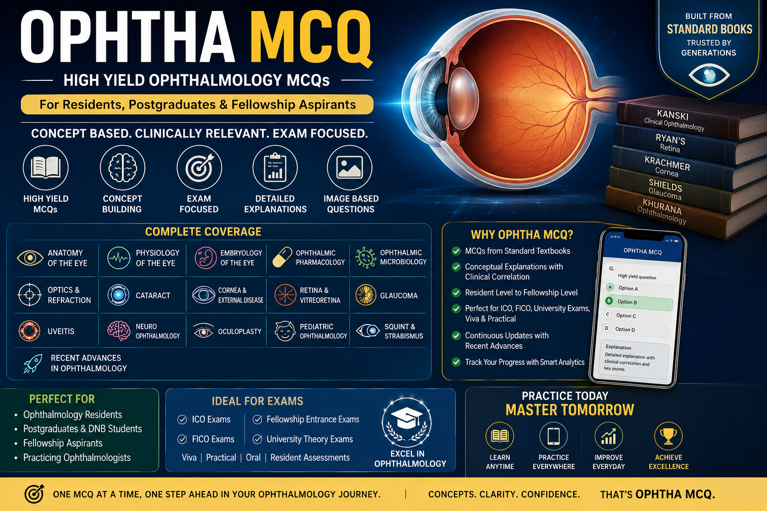
High Yield MCQs
1,000+ high-yield MCQs across all subspecialties with detailed explanations.
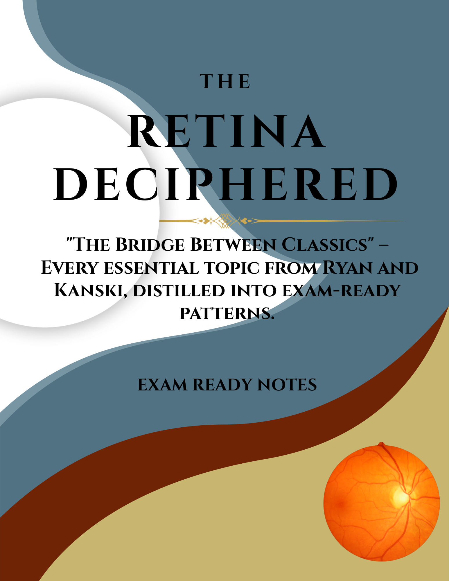
Retina Deciphered
150+ pages of handwritten exam-ready notes covering the entire retina syllabus.

Glaucoma Exam Ready Notes
Complete glaucoma notes from pathophysiology to surgical management.
ICO/FICO Past Papers
Last 10 years of ICO/FICO past papers with full explanations and answer keys.
What's Included
- ✓2000+ mixed ophthalmology MCQs
- ✓Cross-topic explanations
- ✓Combined notes and past papers
- ✓Mobile app access
- ✓Lifetime updates
Covered Topics
- ✓Anatomy
- ✓Optics & Refraction
- ✓Cornea
- ✓Lens & Cataract
- ✓Glaucoma
- ✓Retina
Instant access after purchase. Available on Android & iOS app.
Ophthal MCQs High Yield
High-yield MCQ bank across all ophthalmology subspecialties for rapid revision.
High Yield MCQs
1,000+ high-yield MCQs across all subspecialties with detailed explanations.
Eyelids Exam Ready Notes
Oculoplastics and eyelid disorders for your PG and fellowship exams.
What's Included
- ✓1000+ high-yield MCQs
- ✓Concise explanations
- ✓Quick revision notes
- ✓Mobile app access
- ✓Lifetime updates
Covered Topics
- ✓Cornea
- ✓Glaucoma
- ✓Retina
- ✓Neuro-Ophthalmology
- ✓Uveitis
- ✓Optics & Refraction
Instant access after purchase. Available on Android & iOS app.
Suggested study sequence
Six phases used by recent passers. Spread across 4–8 weeks per subspecialty.
- 1
1. Anatomy and physiology
Layers, innervation, endothelial function, transparency mechanisms.
- 2
2. Infectious keratitis
Bacterial, fungal, viral, acanthamoeba — clinical pattern recognition and treatment.
- 3
3. Dystrophies and degenerations
IC3D classification, genetics, exam appearance.
- 4
4. Ectasia and refractive surgery
Keratoconus, post-LASIK ectasia, CXL, refractive procedures.
- 5
5. Ocular surface disease
Dry eye, MGD, limbal stem cell disease, ocular surface tumours.
- 6
6. Keratoplasty
PK, DALK, DSAEK, DMEK; indications and complications.
Free cornea sample MCQs
A taste of the exam-style stems from the full bank.
Q1.A dendritic ulcer staining with fluorescein in a young adult is most likely caused by:
Explanation: A true branching dendrite with terminal bulbs and ulcerated centre is pathognomonic of HSV epithelial keratitis. HZO produces pseudodendrites without true ulceration.
Q2.The earliest topographic sign of keratoconus is:
Explanation: Asymmetric inferior steepening with skewed radial axes (loss of bowtie symmetry) is the earliest topographic sign.
Q3.Munson's sign is associated with:
Explanation: Munson's sign is V-shaped indentation of the lower eyelid on downgaze due to corneal protrusion in advanced keratoconus.
Cornea FAQ
Answers to the questions residents ask most about this topic.
What is the most common bacterial cause of contact lens keratitis?
When is corneal cross-linking indicated?
Best technique for endothelial keratoplasty?
How do I distinguish granular from macular dystrophy?
Best book for cornea preparation?
Continue your prep
Jump to related exams, topics and resources.
Ready when you are
Your Ophthalmology Exam Is Coming.
Are You Ready?
The world's only ophthalmology PG exam notes and MCQ question bank — built by gold medalists who passed. Start with free sample questions today.